DIAGNOSTIC IMAGING
X-ray
X-ray imaging is a diagnostic procedure that enables us to examine the inside of an animal’s body to assess whether its bones and organs have any problems or diseases. X-ray is a common imaging technique that provides veterinarians with useful information about what is happening inside a dog or a cat.
Ultrasound imaging
Ultrasound imaging is a safe, non-invasive diagnostic procedure that generates a two-dimensional ‘picture’ of your animal’s organs. During the procedure, our expert typically uses sound waves from a hand-held probe to create an image on a monitor.
Magnetic Resonance Imaging (MRI)
“MRI is ideal for detecting and identifying certain cancers. MRI with contrast agents is the best way to examine tumours in the brain, soft tissues, and the spinal cord. Our experts deploy MRI to assess and determine the development stage of cancer, ascertain tumour behaviour, and design and monitor a treatment plan that includes radiotherapy. In addition, MRI provides a non-invasive means to detect benign diseases in animals, including paralyzed dogs with disc herniation, brain tumours, strokes, and cranial cruciate ligament ruptures.
Nuclear Medicine
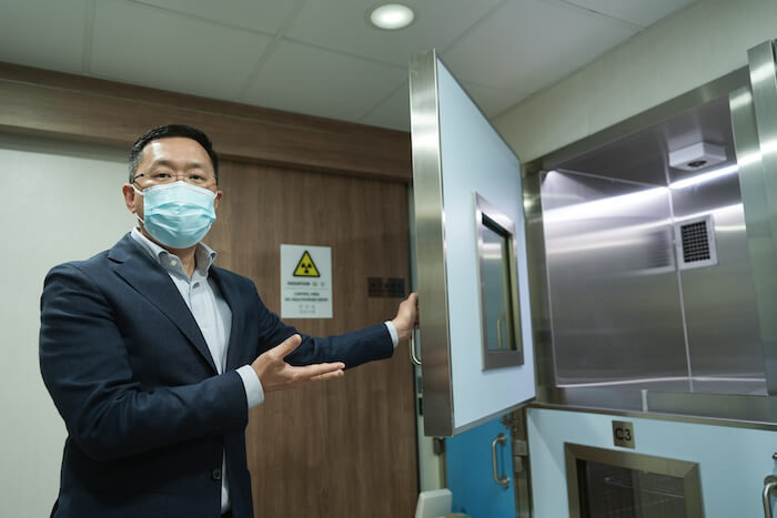
Positron Emission Tomography - Computed Tomography(PET-CT)

Why PET-CT?
PET-CT scanning allows veterinary oncologists to detect changes in the metabolism of cancer cells and to determine whether the cancer has spread to other parts of the body, enabling experts to assess the development stage of the disease. PET-CT scanning can help us to:
- Diagnose cancer
- Determine the size of the cancer and whether it has spread (development stage of cancer)
- Decide whether your animal can have surgery to remove the cancer
- Identify the best treatment for your animal
- Check whether your animal’s cancer has returned
- Plan radiotherapy treatment.
With our oncologists, you can discuss and choose the best options to help your animal and avoid unnecessary treatment.
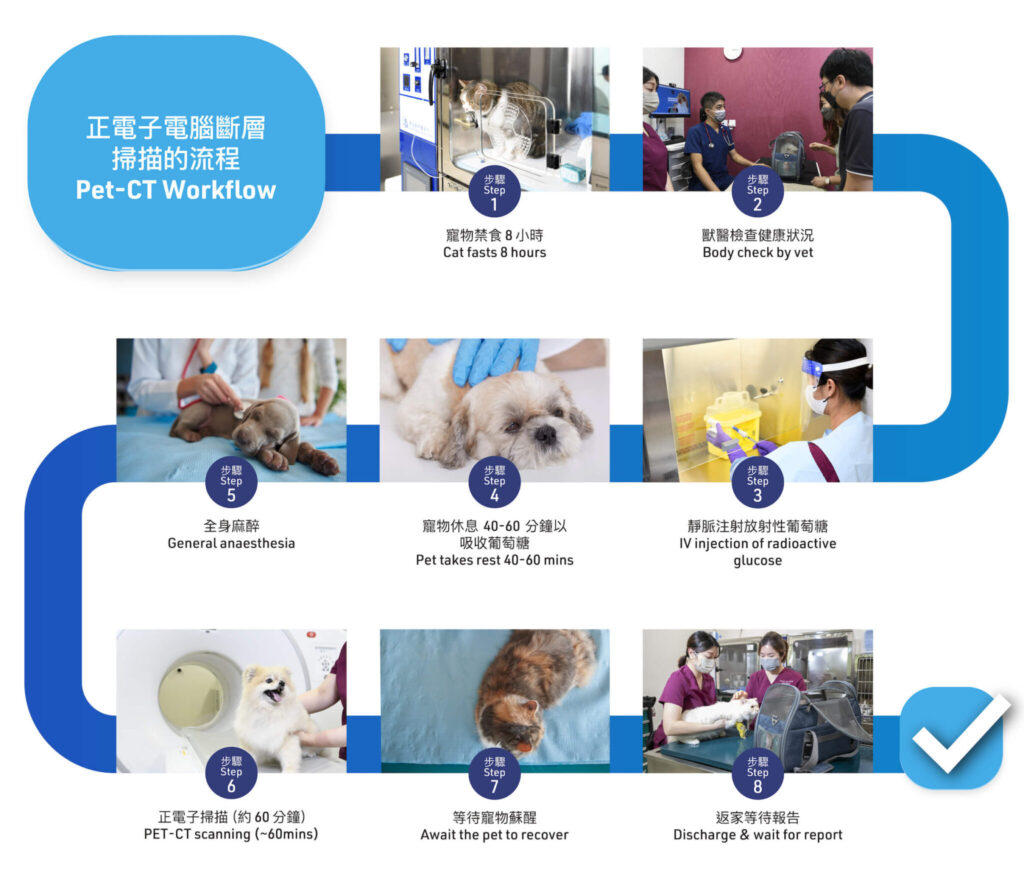
PET-CT Flow chart
Hyperthyroidism in Cats
- Weight loss (despite good appetite).
- Increased appetite and food intake.
- Increased drinking.
- Increased urination.
- Vomiting and/or diarrhoea.
- Coat changes – their coat may appear unkempt, matted or greasy.
I-131 Flow Chart

Home Care After I-131
The owner must sign and retain the provided instruction sheet for post I-131 treatment home care at the time of discharge. Restrictions include minimizing contact with the animal, especially during the first two weeks, handling the cat and waste products with latex gloves for six weeks, and following special waste storage procedures for the first two weeks.